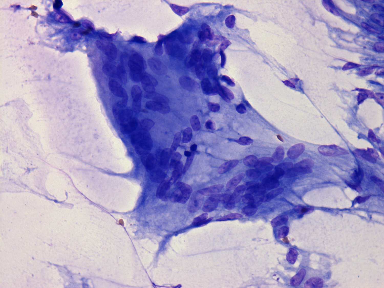
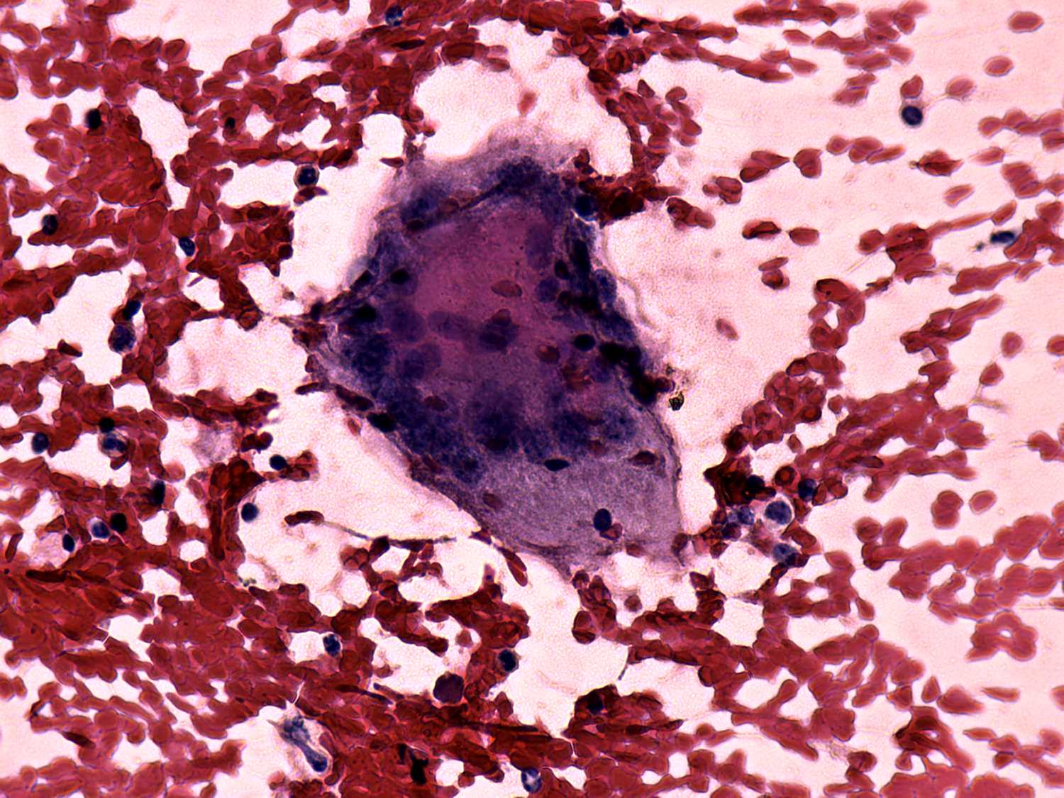
De Quervain's thyroiditis - Figure 3. Cytological presentation.
In the event of a de
Quervain's thyroiditis multinucleated giant cells are in fact part of
the granulation tissue. This is the most important cytological
sign of de Quervain's thyroiditis. Naturally, in a cytological
sample we cannot analyze an intact tissue. Therefore we must keep in
mind that the finding of multinucleated giant cells on a smear itself
is not enough to give the diagnosis of de Quervain's thyroiditis
because benign hyperplastic nodules, papillary carcinoma, Hashimoto's
thyroiditis and granulation around surgical thread may also present
this cell type.
On the other hand, a multinucleated giant cell composed of elongated
epitheloid cells is an almost pathognomic finding. The images in the
first row demonstrate multinucleated cells composed of follicular cell while the images in the second row present multinucleated giant cells
composed of epitheloid cells. The multinucleated cells in the third row
contain both follicular cells and epitheloid cells.
 |
 |
 |
 |
 |
 |